What Is Single Cell Analysis?
Single cell analysis starts with a fundamental shift in perspective: rather than treating tissues as uniform mixtures, it treats each cell as a distinct unit of biology. By analyzing cells one at a time, researchers can detect meaningful differences in gene expression, protein levels, or DNA sequence that bulk methods would miss. This level of detail has revealed previously unrecognized cell types, clarified how cells transition through developmental stages, and helped explain why cells respond differently within the same environment. By revealing this underlying heterogeneity, single cell analysis has become essential for studying complex tissues, understanding disease mechanisms, and identifying novel therapeutic targets. But while it reveals what individual cells are doing, it doesn’t show where they are, or how they’re working together.
Each of the popular single cell analysis techniques listed in the table below offers unique strengths for analyzing cell populations, depending on the biological question at hand.
Single Cell Analysis Technique Strengths and Limitations
| Technique | What It Measures | Strengths | Limitations |
|---|---|---|---|
| Flow Cytometry | Protein markers on or inside cells | Fast, high-throughput, well-established | Limited multiplexing, depends on available antibodies |
| Mass Cytometry (CyTOF) | Protein markers using metal-labeled tags | High multiplexing, minimal spectral overlap | Lower throughput, more complex sample prep |
| scRNA-seq | Gene expression (mRNA) | Comprehensive transcriptomic profiling, unbiased discovery | Requires dissociation, loss of spatial info, may miss rare transcripts |
| scDNA-seq | Genetic variants, copy number, mutations | Single-cell genotyping, useful in cancer and developmental studies | Lower sensitivity, often limited to targeted regions |
With these tools, researchers can uncover rare cell populations, trace developmental lineages, and analyze how individual cells respond to disease or treatment. But these techniques come with trade-offs. Most require that tissues be broken apart into single cells before analysis.
This dissociation step erases the physical organization of the tissue and severs the connections between neighboring cells. You learn who the players are, but not how they interact or where they are positioned. Yet cells do not function in isolation. Their behavior is shaped by their location, their neighbors, and the structural cues of their environment. Without that spatial context, key parts of the biological story remain hidden.
Enter Spatial Omics: Adding Geography to Single Cell Analysis
Spatial omics refers to a group of technologies that combine molecular profiling with spatial information, allowing researchers to see not just what molecules are present in a tissue, but exactly where they are located. These methods reveal insight into how cells are organized, how they communicate, and how their behavior is shaped by their environment by preserving the physical structure of the sample.
Where Spatial Biology Matters Most
Spatial omics turns single-cell data into a map by linking molecular signatures to spatial position, transforming isolated profiles into coordinated patterns that reflect the true complexity of biological systems. This spatial insight is especially critical in areas like:
- Cancer biology
- The location of immune cells within a tumor affects how effectively the body can respond.
- In “cold tumors,” T cells often remain at the edges, unable to access the tumor core.
- Without spatial access, immune responses falter, making tumors more resistant to immunotherapy.
- Knowing which cells are present is important, but knowing where they are is critical.
- Neuroscience
- Neurons follow tightly regulated spatial patterns across brain layers and circuits.
- A neuron’s position often determines its role in signaling or homeostasis.
- Spatial biology helps map these molecular signatures to functional brain architecture.
- Stem cell biology
- Stem cells depend on niche-specific signals to remain quiescent, self-renew, or differentiate.
- When removed from their native microenvironments, their behavior can change dramatically.
- Spatial data reveals how local context shapes stem cell fate and regenerative potential.
How Imaging-Based Spatial Omics Technologies Work
Spatial omics technologies that are imaging-based follow a general workflow that preserves tissue structure while capturing molecular data. Though specific platforms may vary in approach, the core process typically includes four main steps:
- 1. Tissue Sectioning
- Thin slices of tissue are mounted on specialized slides that preserve molecular integrity while maintaining spatial structure.
- Thin slices of tissue are mounted on specialized slides that preserve molecular integrity while maintaining spatial structure.
- 2. Probe Hybridization
- Fluorescent or barcoded probes bind to specific RNA or protein targets, often in multiple rounds to enable high-plex detection.
- 3. High-Resolution Imaging
- Advanced fluorescence microscopy captures both the location and intensity of molecular signals within the tissue.
- 4. Signal Decoding and Spatial Mapping
- Computational tools translate these signals into gene or protein identities and assign them to precise tissue coordinates.
Bridging Modalities for Deeper Insight
While spatial omics offers powerful insights on its own, its value increases even further when paired with complementary technologies. By integrating spatial information with data from other single-cell and molecular approaches, researchers can uncover deeper layers of biological organization and function.
| Modality | How It Combines with Spatial Omics |
|---|---|
| Single-cell RNA sequencing | Defines cell populations by gene expression, which can then be mapped onto tissue to reveal spatial distribution and interactions. |
| Proteomics | Adds protein-level data to RNA maps, helping researchers compare transcription with actual protein localization and abundance. |
| ATAC-seq | Identifies regions of open chromatin, which, when mapped spatially, helps clarify where and how gene regulation is occurring. |
| CRISPR-based screens | Enables targeted gene perturbation, with spatial omics revealing how those changes affect cellular behavior and organization in tissue. |
Discover the Majesty of Spatial Biology
Vizgen’s MERSCOPE Ultra™ platform empowers researchers to explore spatial genomics with unparalleled precision and scalability. By combining high-plex detection of up to 1,000 genes with subcellular resolution, MERSCOPE Ultra™ enables detailed mapping of gene expression across large tissue sections, up to 3.0 cm². Its automated workflow and customizable gene panels make it adaptable for diverse research areas, from neuroscience to oncology.
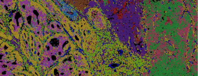
Complementing the platform, Vizgen’s Data Release Program offers a window into what spatial biology actually looks like. These publicly available datasets showcase gene expression mapped at subcellular resolution across diverse tissue types, illustrating how spatial context adds new layers of meaning to single cell analysis. For researchers new to the field or looking to explore specific applications, these examples offer a clear view of what’s possible and what’s next.

Context is Biology
If single cell analysis taught us that no two cells are alike, spatial biology reminds us that no cell acts alone. As technologies continue to illuminate the physical logic of tissues, how structure drives function, how proximity shapes fate, we are moving toward a new way of thinking about biology. Not as a collection of isolated profiles, but as a dynamic system where meaning emerges from context. The challenge now is not just to see more, but to see more clearly, across dimensions, across scales, and across disciplines.

Unlock the Full Potential of Transcriptomics
MERSCOPE UltraTM brings scalability and automation to spatial transcriptomics, delivering high-throughput, single-cell resolution across expansive tissue sections. Its seamless workflow accelerates discovery, making spatial transcriptomic analysis more accessible and efficient for researchers. Unlock deeper biological insights with advanced imaging and multiplexed gene expression.