Having commercially launched the MERSCOPE® Platform in January 2022, we are thrilled to announce the installation of our 100th instrument less than 2 years later. This milestone spatial genomics platform was recently installed in the Single Cell Genomics Core led by Dr. Rui Chen at the Baylor College of Medicine Department of Molecular and Human Genetics in Houston, Texas. (Figure 1)

FIGURE 1: Dr. Rui Chen (left) and Vizgen CEO Terry Lo (right) standing next to the new MERSCOPE instrument in Dr. Chen’s Core laboratory at the Baylor College of Medicine.
The Core at BCM is no stranger to Vizgen’s industry-leading spatial genomics platform, having been one of the earliest adopters of MERSCOPE. The lab utilized one of Vizgen’s Alpha units to create a mouse retina spatial cell atlas at single-cell resolution, going on to publish their studies leveraging this technology in journals such as Nature Communications, Genome Biology, and Cell Genomics. Dr. Chen’s latest research, where his lab profiled over 100,000 cells in the mouse retina, is currently available as a preprint on BioRxiv.
While installing the instrument, Vizgen CEO Terry Lo had the opportunity to sit down with Dr. Chen and discuss his work as well as his thoughts on the impact of spatial genomics on the research community:
Terry: Tell us a bit about the research you and your group are doing here at Baylor College of Medicine
Dr. Chen: We are interested in looking at the relationship between genetic variation and disease, and we’re taking an interdisciplinary approach using innovative methods in genetics and single-cell omics, as well as computational imaging tools to understand the connections.
This is a very big question to address – with over 30 million variants in the human genome, how can we know which ones are important? We use the visual system as a model, and we’re focusing on advancing our ability to identify, assess and predict genetic variants with functional consequences. Our aim is to characterize the genetic factors that underly neural diseases in the visual system, including both inherited and age-related degenerative retinal diseases.
Our highest priorities are the identification of genes and mutations that underly human diseases, the investigation of transcriptomic and epigenetic changes during development and disease at single-cell resolution, and the development of novel therapeutics – including gene therapy, genome editing, and neural regeneration.
Terry: How important is spatial genomics to your work?
Dr. Chen: The great thing about spatial genomics is that it introduces a novel data modality to the omics sphere, enhancing and broadening the scope of existing technologies and opening up a plethora of potential applications capable of addressing a multitude of research questions that spark our interest. These range from understanding tissue patterning and probing cellular interactions to deciphering connections between cells and their microenvironments. I firmly believe that spatial genomics serves as a driving force that will propel our research forward.
Terry: How did you first hear about Vizgen’s MERSCOPE® Platform?
Dr. Chen: As you know, the MERFISH technology that the MERSCOPE Platform uses was developed in the lab of Dr. Xiaowei Zhuang at Harvard University, and since her group published their landmark paper in Science in 2015, I have been closely following the literature on spatial genomic technologies. I formed a collaboration with Dr. Jeffery Moffitt, who is one of the developers of the technology and serves on Vizgen’s Scientific Advisory Board. He was the person who first introduced me to Vizgen and the MERSCOPE Platform.
Terry: What other kinds of technologies are you running at your Core?
Dr. Chen: We offer many services around single-cell omics such as Single-Cell RNA Sequencing (scRNA-seq), Single-cell Assay for Transposase-Accessible Chromatin using Sequencing (scATAC-seq), and Single-Cell Multiomics (scRNA-seq with scATAC-seq). MERFISH is the first – and currently the only – spatial transcriptomics service to be offered at the Core.
Terry: Have you seen any positive changes in your work after the installation and adoption of Vizgen’s spatial genomics platform?
Dr. Chen: Yes, absolutely – the acquisition of this instrument was made possible through a National Institutes of Health (NIH) S10 shared instrument grant and, since we started using it, numerous labs at Baylor College of Medicine have expressed keen interest in this technology. Currently, many of these labs are in the stages of experiment planning and are poised to explore and evaluate this innovative technology in the near future.
Terry: What are some new applications for the MERSCOPE® Platform that you are interested in exploring through your work?
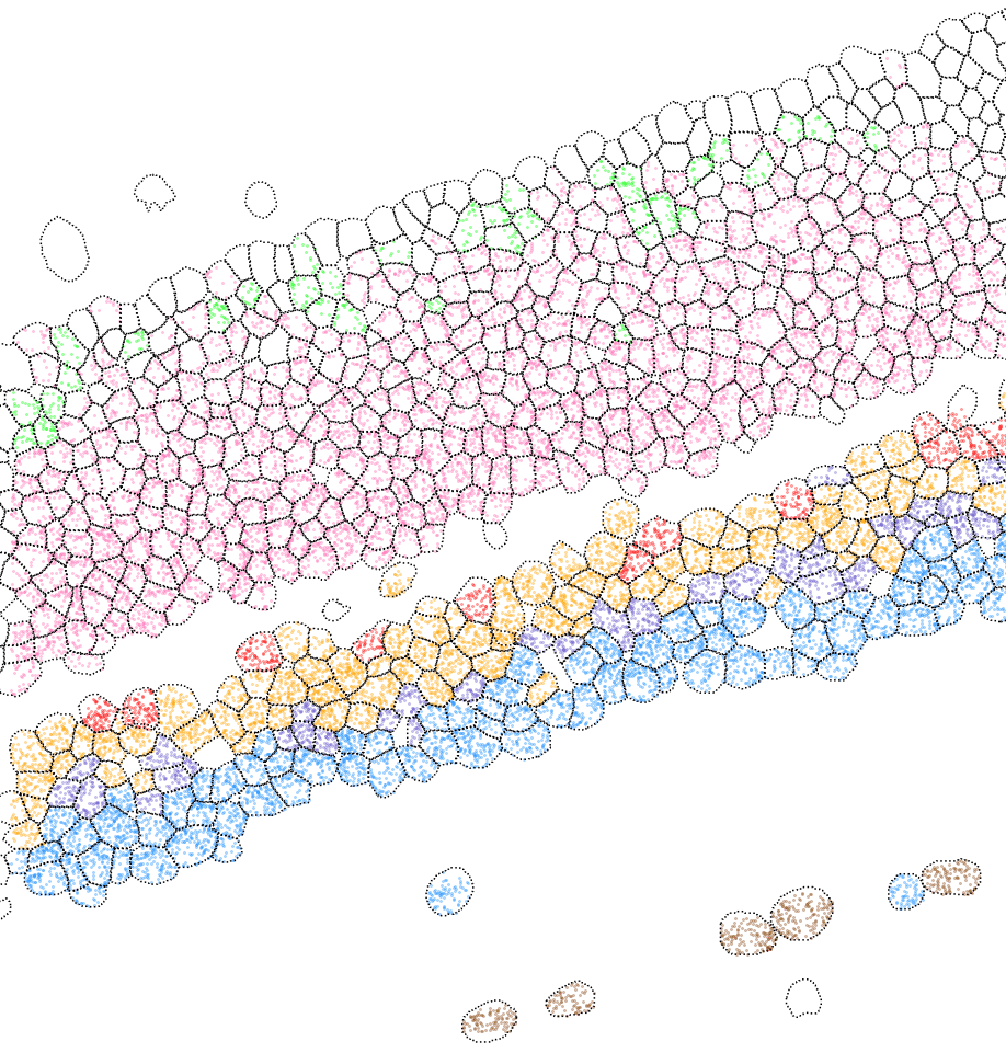
Dr. Chen: We recently completed a project to generate the first ever comprehensive mouse retina spatial cell atlas, using the MERSCOPE Platform to profile over 100,000 cells. The technology enabled us to characterize the spatial distribution of every major retina cell type, with over 100 different subtypes identified. (Figure 2) Not only does this shed light on the structure and development of the mouse retina, but it also now serves as a resource for the scientific community in helping to further research on retinal diseases.


FIGURE 2: The MERSCOPE Platform was used by Dr. Chen’s team to create a mouse retina spatial cell atlas showing the distribution and location of gene expression and cell types across the mouse retina. (A) MERSCOPE Data Image showing the spatial expression of 368 genes across an intact section of the mouse retina. Each colored dot represents an RNA transcript detected by MERSCOPE. (top). (B) MERSCOPE Data Image showing the spatial distribution of major retinal cell types. Pink: Rod, Green: Cone, Gold: BC, Blue: AC, Red: HC, Brown: RGC, Purple: MG. (bottom).
So, aside from cell type identification, we are very interested in applying the MERSCOPE technology to create a spatial map of the human eye, depicting the retina connectivity, combining with Perturb-seq (which uses CRISPR-mediated gene inactivation to assess gene expression phenotypes associated with genetic variation), and characterizing spatial changes during development and disease conditions.
Watch our webinar to learn more about Dr. Chen’s comprehensive mouse retina spatial cell atlas developed with the MERSCOPE spatial genomics platform.